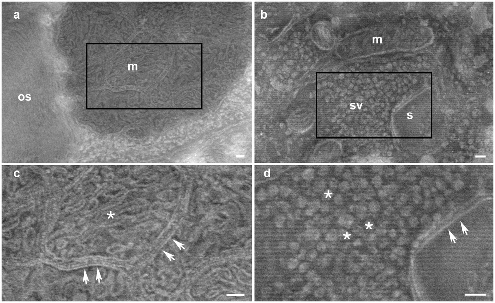
A high magnification image of synapse obtained by electron microscopy
Por um escritor misterioso
Last updated 08 novembro 2024


Molecular self-avoidance in synaptic neurexin complexes

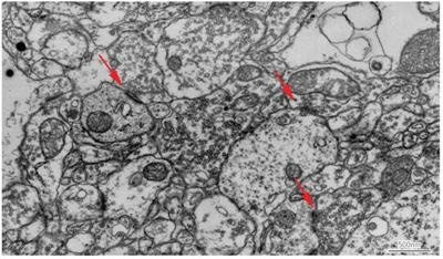
An electron micrograph showing a typical synapse (arrow) within

The 2 Main Electron Microscopy Techniques Explained

Complete Understanding of Structures from Macro to Nanoscales

Synaptic Odyssey Journal of Neuroscience

An electron microscope study of the ring fibers and its

Direct imaging of uncoated biological samples enables correlation

Targeting Functionally Characterized Synaptic Architecture Using

Automated Detection and Localization of Synaptic Vesicles in

Functional Electron Microscopy, “Flash and Freeze,” of Identified
Recomendado para você
-
Steam Workshop::Synapse x08 novembro 2024
-
 Synapse Audio Software Obsession08 novembro 2024
 -
 SynapseX (@SynapseX1) / X08 novembro 2024
 -
 PΛX - Twitter Stats & Analytics HypeAuditor Influencer Marketing Platform08 novembro 2024
 -
 Synapse Wireless Synapse Wireless Presents Facility Performance…08 novembro 2024
 -
 Peer support for people with a brain injury08 novembro 2024
 -
 Synapse Failed To Download Ui Files - Colaboratory08 novembro 2024
 -
 BitAntiCheat - A server-sided, general purpose anti-cheat! - Community Resources - Developer Forum08 novembro 2024
 -
Spaceborne data analysis with Azure Synapse Analytics - Azure Architecture Center08 novembro 2024
-
 family photo (Twitter @gksjenk) link ⏬⏬ i : r/HelluvaBoss08 novembro 2024
você pode gostar
-
 Spy x Family manga: Where to read all chapters right now08 novembro 2024
 -
 Tower Defense 2, Blooket Wiki08 novembro 2024
 -
 Bahamas, el primer gran torneo del 201708 novembro 2024
 -
 Dragon Quest V - HotHB Dragon quest, Dragon warrior, Asian dragon tattoo08 novembro 2024
 -
 Remove Audio Library once and for all or please FIX IT - Website Features - Developer Forum08 novembro 2024
 -
 Palpite Burnley x Tottenham: 02/09/2023 - Campeonato Inglês08 novembro 2024
 -
 Minecraft Tutorial: How to Make Stairs in Minecraft - Howcast08 novembro 2024
 -
 Shadowrun Manual (pdf) :: DJ OldGames08 novembro 2024
 -
 4K wallpapers of Black & Dark in HD, 4K, 5K for PC desktop08 novembro 2024
 -
 Kitchen Sinks, Stylish International Inc.08 novembro 2024

