PDF] Brain Tumor Segmentation of MRI Images Using Processed Image Driven U-Net Architecture
Por um escritor misterioso
Last updated 10 novembro 2024
![PDF] Brain Tumor Segmentation of MRI Images Using Processed Image Driven U-Net Architecture](https://d3i71xaburhd42.cloudfront.net/c750894747d2b3f841de55922b2b68794295de27/7-Table3-1.png)
A fully automatic methodology to handle the task of segmentation of gliomas in pre-operative MRI scans is developed using a U-Net-based deep learning model that reached high-performance accuracy on the BraTS 2018 training, validation, as well as testing dataset. Brain tumor segmentation seeks to separate healthy tissue from tumorous regions. This is an essential step in diagnosis and treatment planning to maximize the likelihood of successful treatment. Magnetic resonance imaging (MRI) provides detailed information about brain tumor anatomy, making it an important tool for effective diagnosis which is requisite to replace the existing manual detection system where patients rely on the skills and expertise of a human. In order to solve this problem, a brain tumor segmentation & detection system is proposed where experiments are tested on the collected BraTS 2018 dataset. This dataset contains four different MRI modalities for each patient as T1, T2, T1Gd, and FLAIR, and as an outcome, a segmented image and ground truth of tumor segmentation, i.e., class label, is provided. A fully automatic methodology to handle the task of segmentation of gliomas in pre-operative MRI scans is developed using a U-Net-based deep learning model. The first step is to transform input image data, which is further processed through various techniques—subset division, narrow object region, category brain slicing, watershed algorithm, and feature scaling was done. All these steps are implied before entering data into the U-Net Deep learning model. The U-Net Deep learning model is used to perform pixel label segmentation on the segment tumor region. The algorithm reached high-performance accuracy on the BraTS 2018 training, validation, as well as testing dataset. The proposed model achieved a dice coefficient of 0.9815, 0.9844, 0.9804, and 0.9954 on the testing dataset for sets HGG-1, HGG-2, HGG-3, and LGG-1, respectively.
![PDF] Brain Tumor Segmentation of MRI Images Using Processed Image Driven U-Net Architecture](https://miro.medium.com/v2/resize:fit:1169/1*Z0Oy_F3W_T5krBslcT6PcQ.png)
Brain Tumor classification and detection from MRI images using CNN based on ResU-Net Architecture, by Sanyukta Suman
![PDF] Brain Tumor Segmentation of MRI Images Using Processed Image Driven U-Net Architecture](https://media.springernature.com/m685/springer-static/image/art%3A10.1038%2Fs41598-023-47107-7/MediaObjects/41598_2023_47107_Fig1_HTML.png)
Utilizing deep learning via the 3D U-net neural network for the delineation of brain stroke lesions in MRI image
![PDF] Brain Tumor Segmentation of MRI Images Using Processed Image Driven U-Net Architecture](https://media.springernature.com/lw685/springer-static/image/art%3A10.1186%2Fs13244-020-00869-4/MediaObjects/13244_2020_869_Fig5_HTML.png)
Convolutional neural networks for brain tumour segmentation, Insights into Imaging
![PDF] Brain Tumor Segmentation of MRI Images Using Processed Image Driven U-Net Architecture](https://file.techscience.com/ueditor/files/cmes/TSP_CMES_128-2/TSP_CMES_14107/TSP_CMES_14107/Images/CMES_14107-fig-5.png/mobile_webp)
MRI Brain Tumor Segmentation Using 3D U-Net with Dense Encoder Blocks and Residual Decoder Blocks
![PDF] Brain Tumor Segmentation of MRI Images Using Processed Image Driven U-Net Architecture](https://media.licdn.com/dms/image/D5612AQHmXP7oVdF7lg/article-cover_image-shrink_600_2000/0/1698339241926?e=2147483647&v=beta&t=KD2ddAt0ARBrWkFWt9qFoICdB7mtpNzg5iOUr3HikdQ)
How U and W Net Architecture in Computer Vision shaped some real work problems in Medical
![PDF] Brain Tumor Segmentation of MRI Images Using Processed Image Driven U-Net Architecture](https://www.mathworks.com/help/images/segment3dbraintumorsusingdeeplearningexample_01_ja_JP.png)
3-D Brain Tumor Segmentation Using Deep Learning - MATLAB & Simulink Example
![PDF] Brain Tumor Segmentation of MRI Images Using Processed Image Driven U-Net Architecture](https://ijisae.org/public/journals/1/submission_2610_2894_coverImage_en_US.png)
Absolute Structure Threshold Segmentation Technique Based Brain Tumor Detection Using Deep Belief Convolution Neural Classifier
![PDF] Brain Tumor Segmentation of MRI Images Using Processed Image Driven U-Net Architecture](https://ijritcc.org/public/journals/1/submission_6457_6403_coverImage_en_US.jpg)
Residual Edge Attention in U-Net for Brain Tumour Segmentation International Journal on Recent and Innovation Trends in Computing and Communication
![PDF] Brain Tumor Segmentation of MRI Images Using Processed Image Driven U-Net Architecture](https://media.springernature.com/m685/springer-static/image/art%3A10.1038%2Fs41598-021-90428-8/MediaObjects/41598_2021_90428_Fig13_HTML.jpg)
Brain tumor segmentation based on deep learning and an attention mechanism using MRI multi-modalities brain images
![PDF] Brain Tumor Segmentation of MRI Images Using Processed Image Driven U-Net Architecture](https://images.prismic.io/encord/57bd343a-7e54-4653-a716-f8fbd88d1afc_image+%284%29.png?auto=compress%2Cformat&fit=max)
Guide to Image Segmentation in Computer Vision: Best Practices
![PDF] Brain Tumor Segmentation of MRI Images Using Processed Image Driven U-Net Architecture](https://pub.mdpi-res.com/computers/computers-10-00139/article_deploy/html/images/computers-10-00139-ag.png?1635497073)
Computers, Free Full-Text
![PDF] Brain Tumor Segmentation of MRI Images Using Processed Image Driven U-Net Architecture](https://www.researchgate.net/publication/344057933/figure/fig1/AS:932932470456320@1599439842224/System-Architecture-Brain-Tumor-Segmentation-using-CNN-is-done-mainly-by-using-three.jpg)
System Architecture Brain Tumor Segmentation using CNN is done mainly
![PDF] Brain Tumor Segmentation of MRI Images Using Processed Image Driven U-Net Architecture](https://journals.sagepub.com/cms/10.1177/20552076221074122/asset/images/large/10.1177_20552076221074122-fig1.jpeg)
Magnetic resonance image-based brain tumour segmentation methods: A systematic review - Jayendra M Bhalodiya, Sarah N Lim Choi Keung, Theodoros N Arvanitis, 2022
![PDF] Brain Tumor Segmentation of MRI Images Using Processed Image Driven U-Net Architecture](https://onlinelibrary.wiley.com/cms/asset/85e7cbd1-a542-4c37-9d7f-16c756eeef98/ima22571-fig-0008-m.jpg)
International Journal of Imaging Systems and Technology, IMA
![PDF] Brain Tumor Segmentation of MRI Images Using Processed Image Driven U-Net Architecture](https://www.frontiersin.org/files/MyHome%20Article%20Library/950706/950706_Thumb_400.jpg)
Frontiers MM-UNet: A multimodality brain tumor segmentation network in MRI images
Recomendado para você
-
 Brain Test Level 191 I hate this! The baby is crying again! Stop this scream in 202310 novembro 2024
 -
 how to win 191 on brain test|TikTok Search10 novembro 2024
 -
 Brain Test: Tricky Puzzles - Seviye 191 • Game Solver10 novembro 2024
 -
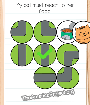 Brain Test 4 Levels 191, 192, 193, 194, 195 Answers10 novembro 2024
 -
 Case 12: left visual'spatial neglect observed in the lime crossing test10 novembro 2024
 -
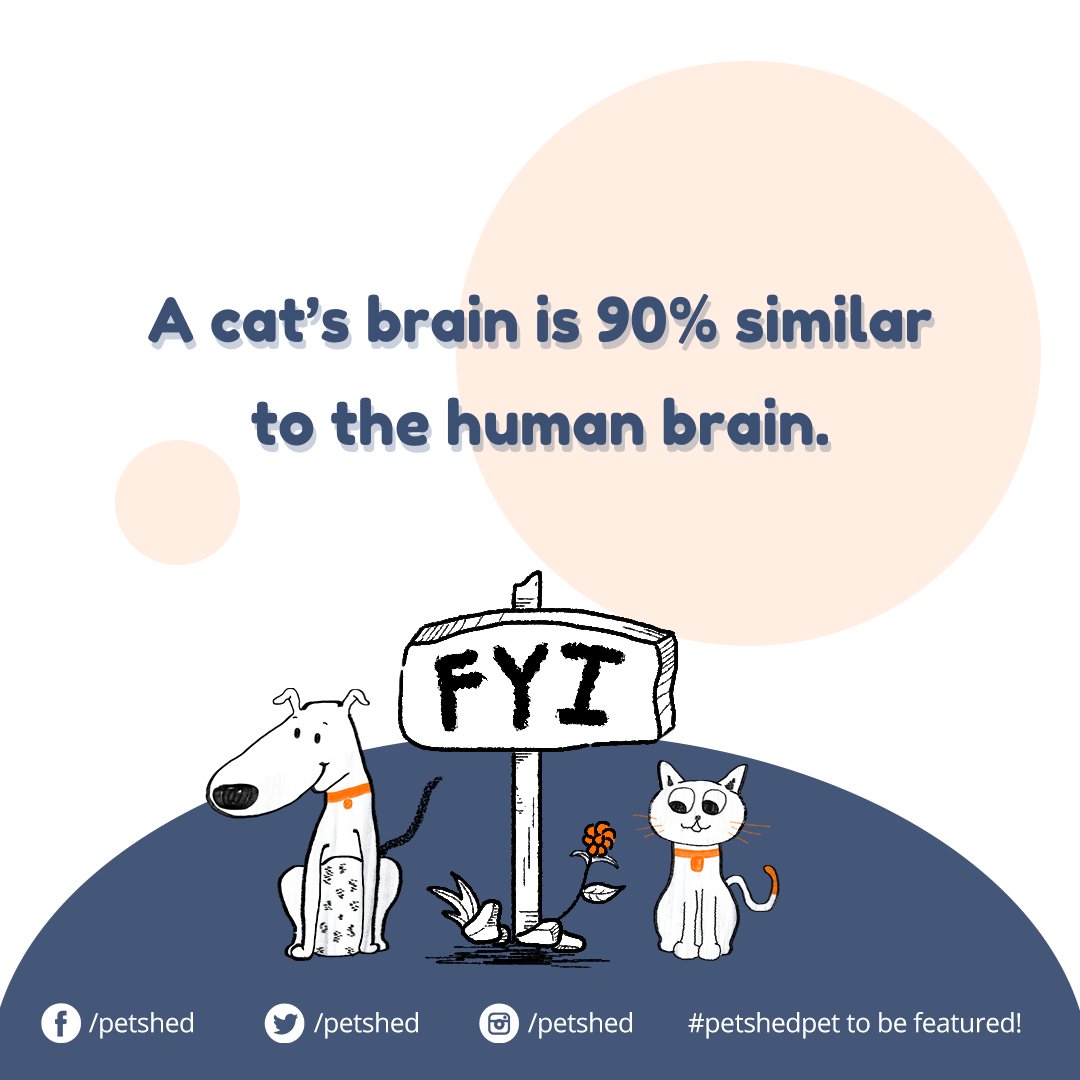 Pet Shed (@PetShed) / X10 novembro 2024
 -
 An Illustrated History of macOS10 novembro 2024
 -
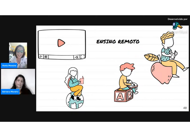 Lapeq10 novembro 2024
 -
 Need Movement Activities To Keep Kids Active At School? - Top Notch Teaching10 novembro 2024
 -
 Brain Test 3 Level 19110 novembro 2024
você pode gostar
-
Boruto solou o Code! Kawaki chegou pra atrapalhar? Boruto Two Blue10 novembro 2024
-
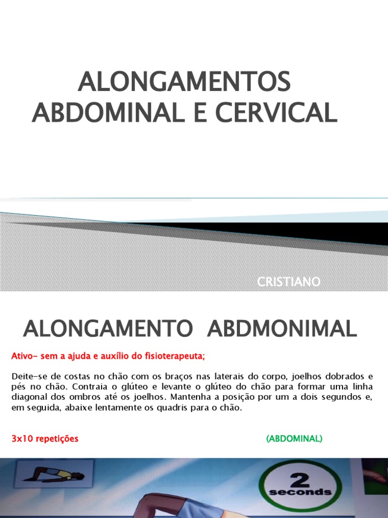
ALONGAMENTOS10 novembro 2024
-
 Chopstick Grips of TWICE Members - Marcosticks10 novembro 2024
 -
:upscale()/2023/02/28/011/n/1922283/tmp_UnERLk_a8945fa65f348ab9_pedro-pascal-bella-ramsey_1.jpg) The Last of Us: Part 2 Game Summary10 novembro 2024
The Last of Us: Part 2 Game Summary10 novembro 2024 -
 Fazendo a Nossa Festa - Colorir: Imagens para Colorir do Chaves e sua Turma!10 novembro 2024
 -
 Watch: Ukrainian Snipers Gun Down Russians, Kyiv It's Close to Breaking World Record10 novembro 2024
 -
 ▻ Assassin's Creed 2 - The Movie All Cutscenes (Full Walkthrough HD)10 novembro 2024
 -
 Médico que receitou videogame e sorvete para criança doente é10 novembro 2024
 -
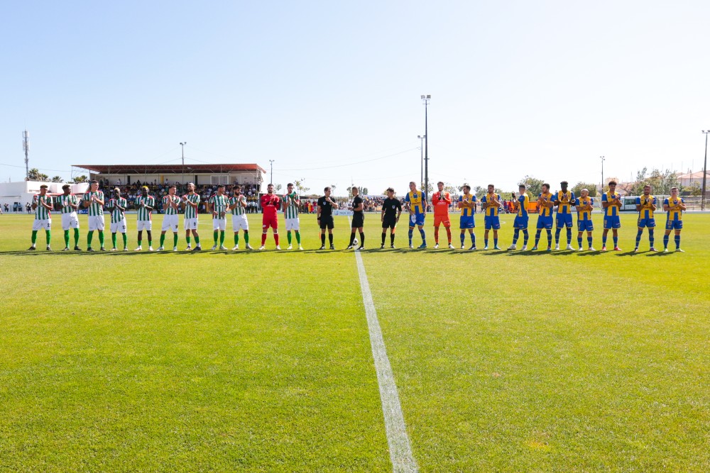 Re)veja a Final da Taça do Algarve esta quarta-feira no Canal 1110 novembro 2024
 -
 انمي Hanma Baki Son of Ogre 2nd Season الحلقة 1 مترجمة اون لاين10 novembro 2024

